By Jaclyn Amaru
Amateur owners, riding professionals, grooms, trainers, barn managers, equine massage therapists, farriers and vets all have one thing in common besides their obvious love for horses. That commonality is that they have all dealt with a horse that is inflamed, under performing, off balance, lame or a combination of these ailments. As we know, horses cannot tell us when something is wrong, so we often do not diagnose these problems until they become obviously evident or after injury has already occurred. But what if there was a technology available that was non-invasive, affordable and sensitive to screen for these hot spots? A tool we could utilize to evaluate for pain management? Equine thermography is taking the industry by storm as more and more professionals become certified to snap the thermal images and interpret their meanings. Here, we will review in lay terms what thermal imaging is and how it is useful.
Thermal imaging has been around for decades, used mainly in military applications. Only until fairly recently were certain spectrums released for public use.
Thermal imaging cameras are specially designed to capture infrared waves and then convert them to an image we can visually interpret. Heat, which is emitted from the skin or just below the skin surface, is generally directly related to increased or poor circulation, or local metabolism, giving rise to hot or cool spots, respectively. Thermal imaging cameras provide snapshots of these physiologic changes related to heat, often the precursor and cardinal sign of injury.
As previously mentioned, application of thermography for equine health is on the rise, but is still widely under utilized. Benefits of thermography include its non-invasive nature, ability to analyze fast with a typical exam ranging from 20-30 minutes, radiation and drug-free testing and usually some degree of on-site interpretation, versus an x-ray or ultrasound which may need to be sent to a radiologist. The pitfall of thermography is that you should be certified as at least a level 1 thermographer by an accredited institution or have attended an equine specific thermography course in order to understand the basics of thermal heat. It is not recommended that you go out and purchase a camera and start shooting pictures, as many factors can influence your results.
To substantiate the use of thermal imaging and its benefits, Dr. Tracy Turner and associate doctors at the University of Minnesota published a study that examined the use of thermal assessment of racing thoroughbreds. This study found that there was a high level of correlation between “trainer-perceived problems and vet diagnoses” with increases in heat evident nearly two weeks before a hot spot became a problem area. Specifically, in every one of the injured horses discovered, their site of injury was evident at least two weeks before diagnosis, while some of the horses that were removed early from the study due to “chronic soreness,” multiple sites of inflammation were discovered as far out as four weeks prior to removal from training. It is said that the thermal imaging camera is some “ten times more powerful than a clinician’s hand.”
As we saw substantiated by a previous study, being able to identify sites of inflammation or poor blood flow could represent an impending injury. Thermal imaging has been used to help detect lameness, hoof balance, laminitis, abscesses, circulatory problems related to navicular syndrome, fractures, bone spurs, back pain, dental problems, etc. Trainers have also been using it to correct saddle fitting and as a pre-purchase requirement on new horses. Not only will a thermal analysis help to prevent injury by identifying a hot spot early, but it will also save you a giant vet bill had the injury actually occurred, provide a baseline to follow thermal patterns, serve as good medical practice for preventative medicine and allow you to make early interventions for pain management, such as stretching, equine massage, acupuncture, or using a tool like Posture Prep to encourage better blood flow, relaxation and reduction of stress and strain to chronically inflamed tissues and ligaments. Generally speaking, the overall cost of a thermal analysis is anywhere from two to four hundred dollars.
The annual thermal imaging conference being held in San Diego, California in September 2016 is dedicating one whole day to an equine specific track, as the requests for training have grown more popular. The conference features teaching sessions, hands-on clinics, saddle fitting basics and getting started running an equine thermography business.
Digital thermal imaging clearly offers an opportunity for horse professionals not already engaged in the practice to expand their business to include a technology that continues to advance and offer insight on the health and well being of our equine counterparts. Not only can it help to identify a problem weeks before a full-blown manifestation, but also serve as a highly effective way to monitor recovery progress or response to change in training programs or sought intervention. It is a wonderful way to actively practice preventative medicine, as well as patrol the welfare of competition. Thermal imaging IS the hot spot.
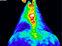
Thermal imaging used to evaluate back
problems.
Here we see a problem later diagnosed as “kissing spine.” This is when a horse feels low grade back pain related to parts of the vertebrae impinging upon each other. Symptoms are often very subtle. Photo credit: http://www.syncequine.com
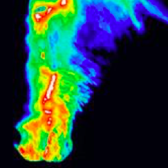
Dental and sinus problems
Dental and sinus problems can also be screened for with thermal imaging. Photo credit: http://www.syncequine.com
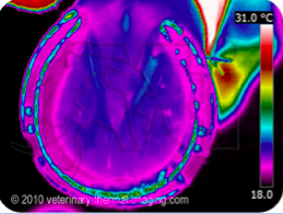
Your horse’s hoof
Checking the balance of your horse’s hoof is important, as well as evaluating the degree of circulation. Poor circulation manifests as a cooler image with non-specific thermal patterns. Photo credit: http://www.veterinary-thermal-imaging.com
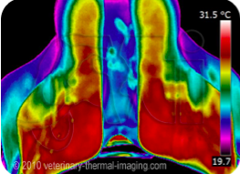
Thermal imaging of a saddle fit
Thermal imaging of a saddle fit which revealed the problem of being too narrow. When a saddle is identified as having the wrong fit, if it is tack that has been in use, it is recommended to follow up with a back imaging consultation. Photo credit:http://www.veterinary-thermal-imaging.com
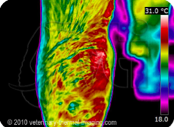
Ligament of the knee
Increased thermal pattern in the right medial collateral ligament of the knee, giving rise to this pony’s waxing and waning lameness. Photo credit: http://www.veterinary-thermal-imaging.com
Sources: www.veterinary-thermal-imaging.com, http://www.thehorse.com/articles/33247/equine-thermography, http://equineir.com/, http://www.unitedinfrared.com/, http://www.flir.com/home/, http://www.infraredtraining.com/, http://www.syncequine.com/, http://docs.fei.org/,

